


sexta-feira, 30 de dezembro de 2011
segunda-feira, 26 de dezembro de 2011

Artigo sobre Microscopia/Documentação - DIGITAL DOCUMENTATION AND THE DENTAL OPERATING MICROSCOPE: WHAT YOU SEE IS WHAT YOU GET
THE INTERNATIONAL JOURNAL OF MICRODENTISTRY
DIGITAL DOCUMENTATION AND THE
DENTAL OPERATING MICROSCOPE:
WHAT YOU SEE IS WHAT YOU GET
Glenn A. van As
The importance of regular digital photography and documentation in the dental
office cannot be understated. The many uses for documentation include
publication, diagnosis and treatment planning, dentolegal documentation,
forensic documentation, insurance verification, patient education, marketing,
and communication with laboratories, dental team members, and colleagues.
With the introduction of the dental operating microscope into dental treatment,
the ease and scope of documentation have been drastically improved. Images
and video taken with the microscope can show details that would not be visible
with conventional photography. The aim of this article is to provide an overview
of the equipment and techniques available to achieve high-quality digital
documentation using an operating microscope.


quinta-feira, 22 de dezembro de 2011
Caso Clínico de Retratamento Endodôntico do dente 26 - Prof. Carlos E. Silveira Bueno
Retratamento Endodôntico do dente 26
Localização do 4o Canal


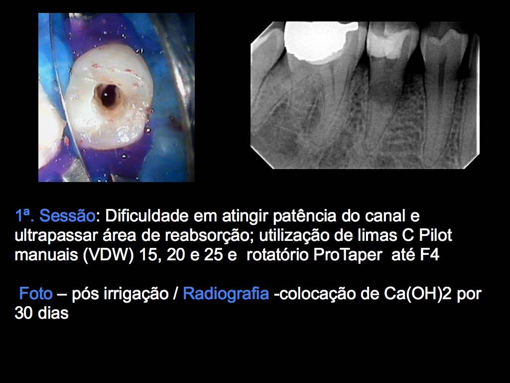
Caso de reabsorção interna/externa - Prof. Carlos E. Silveira Bueno
Caso Clínico de Tratamento de Reabsorção com Vedamento utilizando MTA
Prof. Carlos E. Silveira Bueno


terça-feira, 20 de dezembro de 2011

segunda-feira, 19 de dezembro de 2011
Aula de Acupuntura com a Profa. Fátima Stain
Prof. Fatima


Semana passada os alunos da especialização tiveram a oportunidade de assistir a mais uma aula de Acupuntura voltada a odontologia, aula esta ministrada pela Profa. Fatima Stein.


quarta-feira, 14 de dezembro de 2011
EEC no JOE - Comparison among Manual Instruments and PathFile and Mtwo Rotary Instruments to Create a Glide Path in the Root Canal Preparation of Curved Canals

Comparison among Manual Instruments and PathFile and
Mtwo Rotary Instruments to Create a Glide Path in the Root
Canal Preparation of Curved Canals
Vanessa de Oliveira Alves, DDS, MSc, Carlos Eduardo da Silveira Bueno, DDS, MSc, PhD, Rodrigo Sanches Cunha, DDS, MSc, PhD, S ergio Luiz Pinheiro, DDS, MSc, PhD,
Carlos Eduardo Fontana, DDS, MSc, and Alexandre Sigrist de Martin, DDS, MSc, PhD
sábado, 10 de dezembro de 2011
Motores Elétricos
Bom dia!
Estou para comprar um motor elétrico para instrumentação.
Sö que estou com uma dúvida, será que vale a pena esperar para adquirir um que faça o movimento recíproco tb?
Abs
Obrigado
Eduardo M. Roseffi
(Santos-SP)
Estou para comprar um motor elétrico para instrumentação.
Sö que estou com uma dúvida, será que vale a pena esperar para adquirir um que faça o movimento recíproco tb?
Abs
Obrigado
Eduardo M. Roseffi
(Santos-SP)
quinta-feira, 8 de dezembro de 2011
Casos Clínicos do Aluno de Mestrado em Endodontia - Daniel R. De Assis


Olá.
Envio 2 casos clínicos:
Dente 21 - Instrumentação manual e obturação Técnica Híbrida de Tagger e AH Plus.
Dente 47 - Instrumentação Protaper Universal e obturação Técnica Híbrida de Tagger e AH Plus.
Daniel - Mestrado _ Turma 655/10
Dr. Daniel R. De Assis
Endodontia
Rua Antônio Galvão Cezarino Leite 81, Vila Santa Catarina,
Americana-SP - F.:(19)34618149
Int Endod J dec 2011 - Diagnostic accuracy of limited-volume cone-beam computed tomography in the detection of periapical bone loss: 360 scans versus 180 scans
Diagnostic accuracy of limited-volume cone-beam computed tomography in the detection of periapical bone loss: 360° scans versus 180° scans
S. Lennon1, S. Patel1,2, F. Foschi1, R. Wilson3, J. Davies4, F. Mannocci1
Aim: To investigate the effect of reducing limited-
volume cone-beam computed tomographs arc of rota-
tion from 360° to 180° on the ability to diagnose small,
artificially created apical lesions.
Methodology: Small, artificial apical bone lesions were prepared with a bur in the apical region of the distal root of ten mandibular first molars, in human dry mandibles. The jaws were scanned in a fixed position with limited-volume CBCT making a 360° and 180° arc of rotation, before and after each periapical lesion had been created. A 4 · 4 cm field of view was used at 90 kV, with a current of 4 mA. Ten examiners blinded to the scan parameters and con- trols scored the presence/absence of bone lesions.
Methodology: Small, artificial apical bone lesions were prepared with a bur in the apical region of the distal root of ten mandibular first molars, in human dry mandibles. The jaws were scanned in a fixed position with limited-volume CBCT making a 360° and 180° arc of rotation, before and after each periapical lesion had been created. A 4 · 4 cm field of view was used at 90 kV, with a current of 4 mA. Ten examiners blinded to the scan parameters and con- trols scored the presence/absence of bone lesions.
Intra-examiner reliability was determined after
2weeks, reviewing half the data set. Statistical
analyses with paired t-tests determined the diagnostic
accuracy of the two modalities (360° vs. 180°) in
terms of sensitivity, specificity, receiver operating
characteristic area under the curve, positive predictive
values and negative predictive values.
Results: The mean values for sensitivity of the 360° and 180° scans were 0.91 and 0.89, respectively; their mean specificities were 0.73. No significant differences were reflected in the statistical analyses.
Conclusions: Both 360° and 180° cone-beam com- puted tomography scans yielded similar accuracy in the detection of artificial bone lesions. The use of 180° scans might be advisable to reduce the radiation dose to the patient in line with the ICRP guidance to use as low a dosage as reasonably achievable.
Keywords: 180°, CBCT, periapical bone loss, ROC,
sensitivity, specificity. Results: The mean values for sensitivity of the 360° and 180° scans were 0.91 and 0.89, respectively; their mean specificities were 0.73. No significant differences were reflected in the statistical analyses.
Conclusions: Both 360° and 180° cone-beam com- puted tomography scans yielded similar accuracy in the detection of artificial bone lesions. The use of 180° scans might be advisable to reduce the radiation dose to the patient in line with the ICRP guidance to use as low a dosage as reasonably achievable.
quarta-feira, 7 de dezembro de 2011

segunda-feira, 5 de dezembro de 2011

sábado, 3 de dezembro de 2011
Workshop de Obturação Termoplastica - APCD Jundiaí
Na última quarta-feira dia 30/11 a Equipe de Endodontia de Campinas, representada pelos professores Carlos E. Silveira Bueno e Carlos E. Fontana estiveram na APCD de Jundiaí à convite dos coordenadores Prof. João Velasco e Irineu Polo.
A aula para turma de especialização em Endodontia foi composta de uma parte teórica seguida de um hands-on sobre obturações termoplásticas..


sexta-feira, 2 de dezembro de 2011
JOE dez 2011 - Effect of Canal Length and Curvature on Working Length Alteration with WaveOne Reciprocating Files
Effect of Canal Length and Curvature on Working Length Alteration with WaveOne Reciprocating Files
Elio Berutti, MD, DDS,* Giorgio Chiandussi, MS, PhD,† Davide Salvatore Paolino, MS, PhD,† Nicola Scotti, DDS,* Giuseppe Cantatore, MD,‡ Arnaldo Castellucci, MD, DDS,§
and Damiano Pasqualini, DDS*
and Damiano Pasqualini, DDS*
Introduction: This study evaluated the working length (WL) modification after instrumentation with WaveOne Primary (Dentsply Maillefer, Ballaigues, Switzerland) reciprocating files and the incidence of overinstrumenta- tion in relation to the initial WL. Methods: Thirty-two root canals of permanent teeth were used. The angles of curvature of the canals were calculated on digital radio- graphs. The initial WL with K-files was transferred to the matched WaveOne Primary reciprocating files. After glide paths were established with PathFile (Dentsply Maillefer, Ballaigues, Switzerland), canals were shaped with Wave- One Primary referring to the initial WL. The difference between the postinstrumentation canal length and the initial canal length was analyzed by using a fiberoptic inspection microscope. Data were analyzed with a balanced 2-way factorial analysis of variance (P < .05). Results: Referring to the initial WL, 24 of 32 Wave- One Primary files projected beyond the experimental apical foramen (minimum–maximum, 0.14–0.76 mm). A significant decrease in the canal length after instrumen- tation (95% confidence interval ranging from 0.34 mm to 0.26 mm) was detected. The canal curvature signif- icantly influenced the WL variation (F1 = 30.65, P < .001). The interaction between the initial canal length and the canal curvature was statistically significant (F2 = 4.38, P = .014). Conclusions: Checking the WL before prepa- ration of the apical third of the root canal is recommended when using the new WaveOne NiTi single-file system. (J Endod 2011;37:1687–1690)
Key Words
Nickel-titanium, reciprocating motion, WaveOne, working lengthquinta-feira, 1 de dezembro de 2011
Caso clínico
Boa tarde,
segue anexado tratamento endodôntico do elemento 34 com dois condutos.
Abraço
Eduardo Pastor
Dr. Eduardo A. Casarin Pastor
Cirurgião-Dentista
CRO-SP: 95.131
Rua 7, 192, Centro, Rio Claro/SP
(19) 3534-4748 , (19) 9219-9322
dr.eduardopastor@hotmail.com


Assinar:
Postagens (Atom)


